


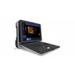
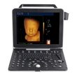
| Monitor: | 15 inch LED medicinal monitor |
| Scanning mode: | convex array/linear array/micro-convex array/phased array |
| Probe interface: | ≥2, double interfaces, probe automatic identification. |
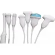
| Support: | convex, linear, trans-vaginal, micro-convex, phased array, 4D volume, rectal probes etc. |
| Language: | Chinese/English/Spanish/French/Russian/Arabic/Vietnamese/Portuguese |
| Display mode: | B mode(B, B+B, 4B, B+M、M), C mode, PW mode, CW mode, real time 3D mode(4D), B/C, B/C/PW, B/PW: 4 scanning speed adjustable |
| Electronic focus: | 4 |
| Body mark: | ≥57 |
| Frequency: | 1.0-18.0MHz multi-frequency |
| Convex probe frequency range: | 1.0, 2.0, 3.0, 3.5, 4.0, 5.5, 6.5, 7.0MHz |
| Trans-vaginal probe frequency range: | 5.0, 6.0, 6.5, 7.5, 9.0MHz |
| Phased array probe (adult) frequency range: | 2.1, 3.0, 3.5, 4.0, 5.0MHz |
| Phased array probe (pediatric) frequency range: | 4.0, 5.5, 6.5, 7.5, 10.0MHz |
| Linear probe frequency range: | 6.0, 6.5, 7.5, 10.0, 12.0, 14.0, 16.0, 18.0MHz |
| Micro-convex probe frequency range: | 2.0, 3.0, 3.5, 4.0, 5.5, 6.5, 7.5, 10.0MHz |
| Rectal probe frequency range: | 6.0, 6.5, 7.5, 10.0, 12.MHz |
| 4D volume probe frequency: | 2.0, 3.0, 3.5, 4.0, 5.5MHz |
| Image processing: | up/down、left/right、angle、blood flow reversal |
| Magnification: | 2-10 times magnification |
| Automatic freeze function: | effective protection of probe life without any operation |
| Alternative image storage method: | UI storage and image storage only,Cine loop speed, visible and adjustable |
| Measurement: | distance, circumference, area, volume, heart rate, tube diameter, narrow rate, angle, speed, abdomen, cardiac, general, musculoskeletal, early obstetric, middle and late obstetric, pelvic(uterus accessory), small organ, urology, peripheral, gestational week and EDD, fetal weight, etc, formula can be converted to edit in obstetric measurement. The distance, circumference and volume measurement of 3D mode. |
| Note: | date, clock, name, gender, age, doctor, hospital, note (full screen character editing)Cine loop≥1200 frames, can be viewed continuously or by frame |
| Storage: | in the storage display interface can be sent to directly to the mobile device, image storage path (can be stored directly to the U disk and other external storage devices), probe parameter storage, cine loop storage, measurement results storage, report storage. |
| Gray Scale: | 256 |
| Dynamic range: | 0-390dB |
| Intelligent TGC: | 8 segments |
| Pre-processiong: | variable aperture, dynamic apodization, dynamic digital filtering, multi-beam parallel processing technology, THI etc. |
Post-processing: | Dynamic range 0-390dB, black and white afterglow 0-7 Smoothing 0-7 Gray curve 1-16 Frame correlation SHG (Second Harmonic Generation) Sound power Wall filter Cumulative number, baseline adjustment, sampling frame adjustment, spectrum sampling volume, spectral sampling volume angle, PRF, etc. |
| Blind zone: | ≤4 |
| Max scanning depth: | 390mm |
| Geometric accuracy: | horizontal≤5%, vertical≤5% |
| Resolution: | lateral≤2mm, axial≤1mm |
| External interface: | DP, HDMI, LAN *2, USB *4, Audio |
| Magnification: | 16 kinds; disease diagnosis more accurate |
| Frame rate: | 15-1016fps (adjustable as per requirement) |
| Scanning range: | 5%--100% |
| Gain control: | Total gain 0~127dB, PW gain 0-15, CFM gain: 0-15 |
| Image optimization: | 6 level adjustable |
| Smoothing: | 8 level adjustable |
| Edge enhancement: | 8 level adjustable |
| PRF: | 16 level adjustable (1.82-29.43KHz) |
| Gray level curve: | 16 level adjustable |
| Sound power: | 15 level adjustable |